

Lattice Degeneration
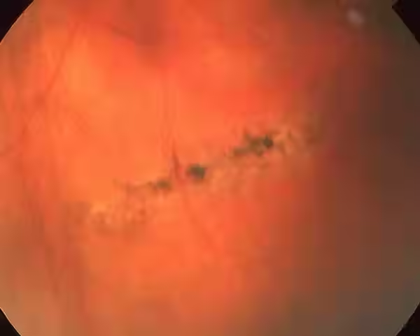
Lattice degeneration is a peripheral retinal change seen on exam in which certain areas of the retina become thin, white, and crisscrossed in a lattice-like pattern.
What Is Lattice Degeneration?
Lattice degeneration is a peripheral retinal change seen on exam in which certain areas of the retina become thin, white, and crisscrossed in a lattice-like pattern. These areas may also develop small atrophic holes. It is a common degenerative finding in the retina and is usually discovered during routine dilated eye exams.
Who it affects:
It can occur in many people and is more likely in individuals with myopia (nearsightedness). Lattice changes may be found in one or both eyes.
Common Symptoms
Most people with lattice degeneration do not have any symptoms. However, if the retina develops a tear or a retinal detachment in the area of the lattice, symptoms may include:
Flashes of light
New floaters
A shadow or curtain in peripheral vision

Treatments
Lattice degeneration itself usually does not require treatment if it’s stable and without tears. However, if associated with retinal holes or early detachment, treatment such as laser retinopexy may be recommended to help “wall off” the area and reduce risk of retinal detachment.
Early diagnosis and treatment can improve your long-term visual outcome. Contact us today for a consultation.
.png)
Frequently Asked Questions

Real Patient Experiences

Bear Findley
2026-03-10
Excellent retina specialist! So happy I found Dr Wong! 👏👏👏👏👏

Gabriela Rossello
2026-03-08
Dr. Wong is very nice, efficient and thorough. She explained everything to me as she was doing the exam. Went over findings. Made recommendations and a plan of action. It is great that there is free parking. Her office is very nice and spacious. For my first time ever visiting an ophthalmologist, this was a very good experience. I highly recommend her.

Robin Frank
2026-02-26
Dr. Wong is an outstanding retina specialist in Bethesda, Maryland. She is incredibly skilled, attentive, and thorough in her evaluations. She carefully explains diagnoses and treatment options, ensuring I feel informed and supported. Her bedside manner is excellent — she is patient, kind, and truly listens. The office is well-organized, and the overall experience is always positive. I highly recommend Dr. Wong to anyone seeking top-quality retinal care. Ms. R.F.

mamet amantai191615
2026-02-17
Good service and good treatment methods

flyhigh72 “flyhigh”
2026-01-23
Dr. Wong is a very thorough and knowledgeable physician. Our visit was extremely pleasant, and she took the time to provide clear and detailed explanations. The front desk staff were exceptionally friendly and helpful, and the office is beautifully decorated and very comfortable. Overall, it was a wonderful and pleasantly surprising experience for us.
4.5/5
Rating
Based on
4.5/5
based on
Rating